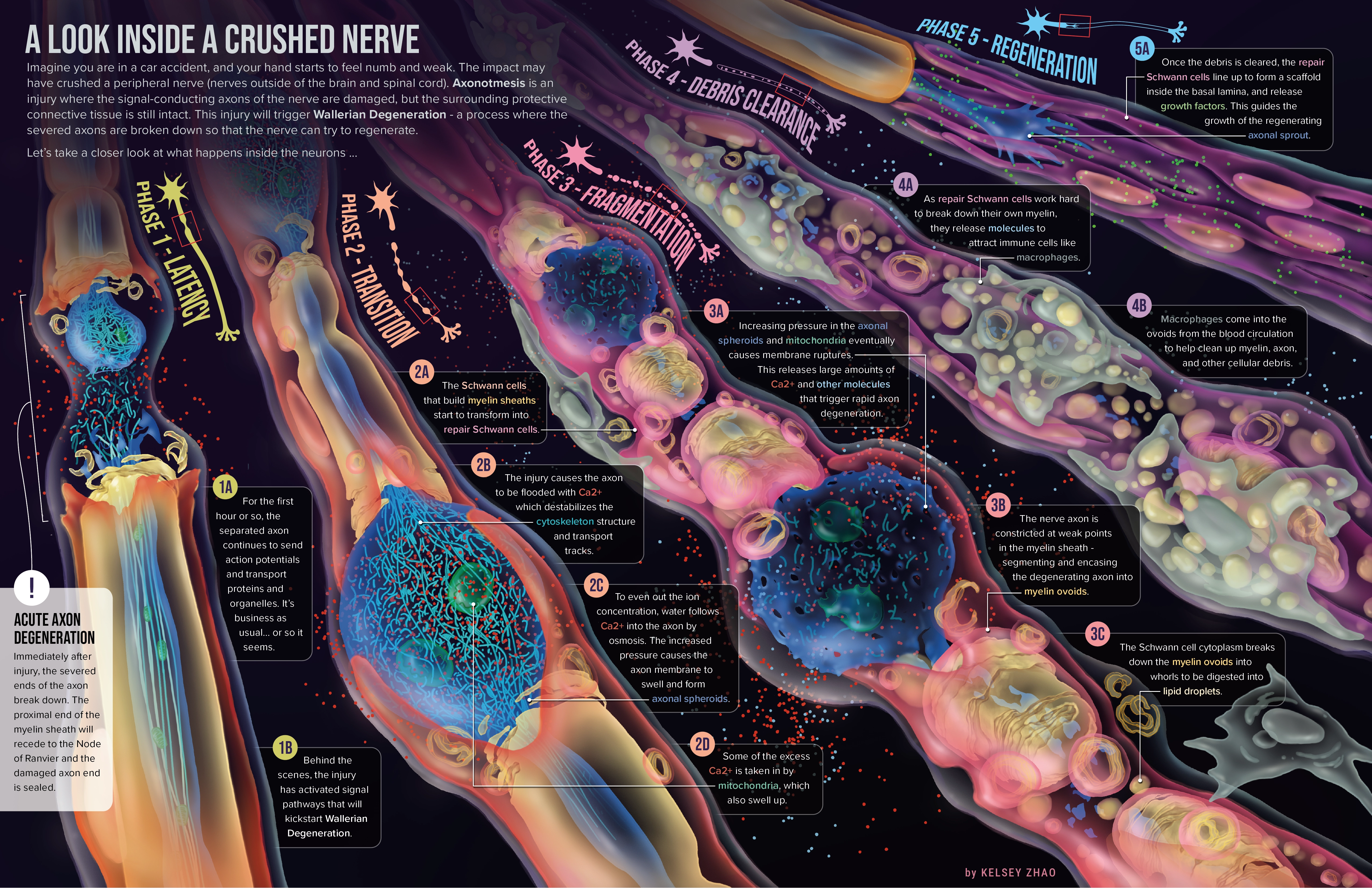
This conceptual medical illustration describes the spatial and cellular changes in the distal axon of a peripheral
nerve after an axonal crush injury. The stages of nerve and axon degeneration are illustrated
chronologically from left to right, with small diagrams to orient the reader to the featured axon region.
The text is colour-coded with the illustrations to describe the specific cellular and molecular processes
that bring about this transformation. The visual design of this piece
takes inspiration from sci-fi art to evoke a sense of wonder and awe towards the intricate processes happening in
our bodies every day.
References
Barrientos, S. A., Martinez, N. W., Yoo, S., Jara, J. S., Zamorano, S., Hetz, C., Twiss, J. L., Alvarez, J., & Court, F. A. (2011). Axonal Degeneration Is Mediated by the Mitochondrial Permeability Transition Pore. The Journal of Neuroscience, 31(3), 966–978. https://doi.org/10.1523/JNEUROSCI.4065-10.2011
Beirowski, B., Nógrádi, A., Babetto, E., Garcia-Alias, G., & Coleman, M. P. (2010). Mechanisms of Axonal Spheroid Formation in Central Nervous System Wallerian Degeneration. Journal of Neuropathology and Experimental Neurology, 69(5), 455–472. https://doi.org/10.1097/NEN.0b013e3181da84db
Catenaccio, A., Llavero Hurtado, M., Diaz, P., Lamont, D. J., Wishart, T. M., & Court, F. A. (2017). Molecular analysis of axonal-intrinsic and glial-associated co-regulation of axon degeneration. Cell Death & Disease, 8(11), Article e3166. https://doi.org/10.1038/cddis.2017.489
Datar, A., Ameeramja, J., Bhat, A., Srivastava, R., Mishra, A., Bernal, R., Prost, J., Callan-Jones, A., & Pullarkat, P. A. (2019). The Roles of Microtubules and Membrane Tension in Axonal Beading, Retraction, and Atrophy. Biophysical Journal, 117(5), 880–891. https://doi.org/10.1016/j.bpj.2019.07.046
Eddleman, C. S., Bittner, G. D., & Fishman, H. M. (2000). Barrier Permeability at Cut Axonal Ends Progressively Decreases until an Ionic Seal Is Formed. Biophysical Journal, 79(4), 1883–1890. https://doi.org/10.1016/S0006-3495(00)76438-1
Faris, H., Almasieh, M., & Levin, L. A. (2021). Axonal degeneration induces distinct patterns of phosphatidylserine and phosphatidylethanolamine externalization. Cell Death Discovery, 7(1), Article 247. https://doi.org/10.1038/s41420-021-00641-7
Gomez-Sanchez, J. A., Carty, L., Iruarrizaga-Lejarreta, M., Palomo-Irigoyen, M., Varela-Rey, M., Griffith, M., Hantke, J., Macias-Camara, N., Azkargorta, M., Aurrekoetxea, I., De Juan, V. G., Jefferies, H. B. J., Aspichueta, P., Elortza, F., Aransay, A. M., Martínez-Chantar, M. L., Baas, F., Mato, J. M., Mirsky, R., … Jessen, K. R. (2015). Schwann cell autophagy, myelinophagy, initiates myelin clearance from injured nerves. The Journal of Cell Biology, 210(1), 153–168. https://doi.org/10.1083/jcb.201503019
Gomez-Sanchez, J. A., Pilch, K. S., van der Lans, M., Fazal, S. V., Benito, C., Wagstaff, L. J., Mirsky, R., & Jessen, K. R. (2017). After Nerve Injury, Lineage Tracing Shows That Myelin and Remak Schwann Cells Elongate Extensively and Branch to Form Repair Schwann Cells, Which Shorten Radically on Remyelination. The Journal of Neuroscience, 37(37), 9086–9099. https://doi.org/10.1523/JNEUROSCI.1453-17.2017
Hall, W. A., Nadi, M. (2023). Axonotmesis. In StatPearls [Internet]. StatPearls Publishing. https://www.ncbi.nlm.nih.gov/books/NBK482369/
Hunter-Chang, S., Kim-Aun, C., Karim, H., Jones, M., Vegiraju, T., Stepanova, E., Manke, B., Kucenas, S., & Deppmann, C. (2025). Axonal spheroids are regulated by Schwann cells after peripheral nerve injury. Glial Health Research, 2, Article 100005. https://doi.org/10.1016/j.ghres.2025.100005
Jang, S. Y., Shin, Y. K., Park, S. Y., Park, J. Y., Lee, H. J., Yoo, Y. H., Kim, J. K., & Park, H. T. (2016). Autophagic myelin destruction by schwann cells during wallerian degeneration and segmental demyelination. Glia, 64(5), 730–742. https://doi.org/10.1002/glia.22957
Jessen, K. R., & Mirsky, R. (2016). The repair Schwann cell and its function in regenerating nerves. The Journal of Physiology, 594(13), 3521–3531. https://doi.org/10.1113/JP270874
Jo, Y. R., Oh, Y., Kim, Y. H., Shin, Y. K., Kim, H. R., Go, H., Shin, J., Park, H. J., Koh, H., Kim, J. K., Shin, J. E., Lee, K. E., & Park, H. T. (2023). Adaptive autophagy reprogramming in Schwann cells during peripheral demyelination. Cellular and Molecular Life Sciences : CMLS, 80(1), Article 34. https://doi.org/10.1007/s00018-022-04683-7
Ko, K. W., Devault, L., Sasaki, Y., Milbrandt, J., & DiAntonio, A. (2021). Live imaging reveals the cellular events downstream of SARM1 activation. eLife, 10, Article 71148. https://doi.org/10.7554/eLife.71148
Napoli, I., Noon, L. A., Ribeiro, S., Kerai, A. P., Parrinello, S., Rosenberg, L. H., Collins, M. J., Harrisingh, M. C., White, I. J., Woodhoo, A., & Lloyd, A. C. (2012). A Central Role for the ERK-Signaling Pathway in Controlling Schwann Cell Plasticity and Peripheral Nerve Regeneration In Vivo. Neuron (Cambridge, Mass.), 73(4), 729–742. https://doi.org/10.1016/j.neuron.2011.11.031
Mansoor Rayegani, S. (2012). Basic principles of peripheral nerve disorders (S. M. Rayegani, Ed.). IntechOpen.
Shacham-Silverberg, V., Sar Shalom, H., Goldner, R., Golan-Vaishenker, Y., Gurwicz, N., Gokhman, I., & Yaron, A. (2018). Phosphatidylserine is a marker for axonal debris engulfment but its exposure can be decoupled from degeneration. Cell Death & Disease, 9(11), Article 1116. https://doi.org/10.1038/s41419-018-1155-z
Stoll, G., Griffin, J. W., Li, C. Y., & Trapp, B. D. (1989). Wallerian degeneration in the peripheral nervous system: participation of both Schwann cells and macrophages in myelin degradation. Journal of Neurocytology, 18(5), 671–683. https://doi.org/10.1007/BF01187086
Tomatsuri, M., Okajima, S., & Ide, C. (1993). Sprout formation at nodes of Ranvier of crush-injured peripheral nerves. Restorative Neurology and Neuroscience, 5(4), 275–282. https://doi.org/10.3233/rnn-1993-5403
Yong, Y., Hunter-Chang, S., Stepanova, E., & Deppmann, C. (2021). Axonal spheroids in neurodegeneration. Molecular and Cellular Neuroscience, 117, Article 103679. https://doi.org/10.1016/j.mcn.2021.103679
Yong, Y., Gamage, K., Cheng, I., Barford, K., Spano, A., Winckler, B., & Deppmann, C. (2019). p75NTR and DR6 Regulate Distinct Phases of Axon Degeneration Demarcated by Spheroid Rupture. The Journal of Neuroscience, 39(48), 9503–9520. https://doi.org/10.1523/JNEUROSCI.1867-19.2019
Zhang, K., Jiang, M., & Fang, Y. (2021). The Drama of Wallerian Degeneration: The Cast, Crew, and Script. Annual Review of Genetics, 55(1), 93–113. https://doi.org/10.1146/annurev-genet-071819-103917
Zochodne, D. W. (2000). The microenvironment of injured and regenerating peripheral nerves. Muscle & Nerve, 23(S9), S33–S38. https://doi.org/10.1002/1097-4598(2000)999:9<::AID-MUS7>3.0.CO;2-F